Anti-Glucose transporter 2 Antibody (56573)
$466.00
SKU: 56573
Categories: Antibody Products, Neuroscience and Signal Transduction Antibodies, Products
Overview
Product Name Anti-Glucose transporter 2 Antibody (56573)
Description Anti-Glucose transporter 2 Rabbit Polyclonal Antibody
Target Glucose transporter 2
Species Reactivity Human, Rat
Applications WB,ICC/IF
Host Rabbit
Clonality Polyclonal
Immunogen Synthetic peptide corresponding to the C-terminus of human GLUT2.
Properties
Form Liquid
Concentration 1.0 mg/mL
Formulation PBS, pH 7.4, 50% glycerol, 0.09% sodium azide.
Buffer Formulation Phosphate Buffered Saline
Buffer pH pH 7.4
Buffer Anti-Microbial 0.09% Sodium Azide
Buffer Cryopreservative 50% Glycerol
Format Purified
Purification Purified by peptide immuno-affinity chromatography
Specificity Information
Specificity This antibody recognizes human and rat GLUT2..
Target Name Solute carrier family 2, facilitated glucose transporter member 2
Target ID Glucose transporter 2
Uniprot ID P11168
Alternative Names Glucose transporter type 2, liver, GLUT-2
Gene Name SLC2A2
Sequence Location Cell membrane, Multi-pass membrane protein
Biological Function Facilitative hexose transporter that mediates the transport of glucose and fructose (PubMed:8027028, PubMed:16186102, PubMed:23396969, PubMed:28083649). Likely mediates the bidirectional transfer of glucose across the plasma membrane of hepatocytes and is responsible for uptake of glucose by the beta cells; may comprise part of the glucose-sensing mechanism of the beta cell (PubMed:8027028). May also participate with the Na(+)/glucose cotransporter in the transcellular transport of glucose in the small intestine and kidney (PubMed:3399500). Also able to mediate the transport of dehydroascorbate (PubMed:23396969). {PubMed:16186102, PubMed:23396969, PubMed:28083649, PubMed:3399500, PubMed:8027028}.
Research Areas Neuroscience
Background GLUT2 is a member of the human glucose transport protein family. It is present in high concentration in the plasma membrane of pancreatic b-cells where it initiates the first step in glucose-stimulated insulin secretion. GLUT2 is required for glucose sensitivity in the hypothalamus and brain stem and is involved in control of food intake and stimulation of glucose uptake by peripheral tissues.
Application Images


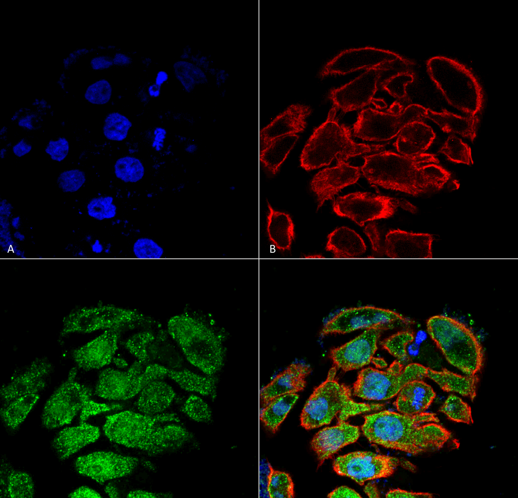
Description Immunocytochemistry/Immunofluorescence analysis using Rabbit Anti-GLUT2 Polyclonal Antibody (56573). Tissue: Colon cancer cell line (HT-29). Species: Human. Fixation: 4% Formaldehyde for 15 min at RT. Primary Antibody: Rabbit Anti-GLUT2 Polyclonal Antibody (56573) at 1:100 for 60 min at RT. Secondary Antibody: Goat Anti-Rabbit ATTO 488 at 1:100 for 60 min at RT. Counterstain: Phalloidin Texas Red F-Actin stain; DAPI (blue) nuclear stain at 1:1000, 1:5000 for 60min RT, 5min RT. Localization: Cytoplasm, membrane. Magnification: 60X. (A) DAPI (blue) nuclear stain (B) Phalloidin Texas Red F-Actin stain (C) GLUT2 Antibody (D) Composite.

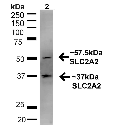
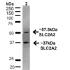
Description Western blot analysis of Rat Liver showing detection of ~57.5kDa GLUT2 protein using Rabbit Anti-GLUT2 Polyclonal Antibody (56573). Lane 1: MW Ladder. Lane 2: Rat Liver (20 µg). Load: 20 µg. Block: 5% milk + TBST for 1 hour at RT. Primary Antibody: Rabbit Anti-GLUT2 Polyclonal Antibody (56573) at 1:1000 for 1 hour at RT. Secondary Antibody: Goat Anti-Rabbit: HRP at 1:2000 for 1 hour at RT. Color Development: TMB solution for 12 min at RT. Predicted/Observed Size: ~57.5kDa. Other Band(s): ~37kDa.

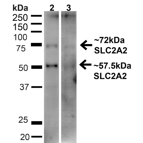
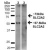
Description Western blot analysis of Human HeLa and HEK293T cell lysates showing detection of ~57.5kDa GLUT2 protein using Rabbit Anti-GLUT2 Polyclonal Antibody (56573). Lane 1: MW Ladder. Lane 2: Human HeLa (20 µg). Lane 3: Human 293T (20 µg). Load: 20 µg. Block: 5% milk + TBST for 1 hour at RT. Primary Antibody: Rabbit Anti-GLUT2 Polyclonal Antibody (56573) at 1:1000 for 1 hour at RT. Secondary Antibody: Goat Anti-Rabbit: HRP at 1:2000 for 1 hour at RT. Color Development: TMB solution for 12 min at RT. Predicted/Observed Size: ~57.5kDa. Other Band(s): ~72kDa.
Handling
Storage This antibody is stable for at least one (1) year at -20°C.
Dilution Instructions Dilute in PBS or medium that is identical to that used in the assay system.
Application Instructions Immunoblotting: use at dilution of 1:1,000. A bands of 57~kDa is detected. Bands of ~72kDa and ~37kDa may also be detected.
Immunofluorescence: use at dilution of 1:100
These are recommended working dilutions.
Endusers should determine optimal dilutions for their applications.
Immunofluorescence: use at dilution of 1:100
These are recommended working dilutions.
Endusers should determine optimal dilutions for their applications.
References & Data Sheet
Data Sheet  Download PDF Data Sheet
 Download PDF Data Sheet

