Anti-CLAC-P Antibody (28115)
$397.00
SKU: 28115
Categories: Antibody Products, Neuroscience and Signal Transduction Antibodies, Products
Overview
Product Name Anti-CLAC-P Antibody (28115)
Description Anti-CLAC-P Rabbit Polyclonal Antibody
Target CLAC-P
Species Reactivity Human, Mouse
Applications WB,IHC
Host Rabbit
Clonality Polyclonal
Immunogen Synthetic peptide corresponding to aa 430-445 of the NC3 region of human and mouse CLAC-P.
Properties
Form Liquid
Concentration Lot Specific
Formulation PBS, pH 7.4.
Buffer Formulation Phosphate Buffered Saline
Buffer pH pH 7.4
Format Purified
Purification Purified by immunoaffinity chromatography
Specificity Information
Specificity This antibody recognizes native and human and mouse CLAC-P.
Target Name Collagenα-1(XXV) chain
Target ID CLAC-P
Uniprot ID Q9BXS0
Alternative Names Alzheimer disease amyloid-associated protein, AMY, CLAC-P [Cleaved into: Collagen-like Alzheimer amyloid plaque component
Gene Name COL25A1
Sequence Location Membrane
Biological Function Inhibits fibrillization of amyloid-beta peptide during the elongation phase. Has also been shown to assemble amyloid fibrils into protease-resistant aggregates. Binds heparin. {PubMed:15522881, PubMed:15615705, PubMed:15853808, PubMed:16300410}.
Research Areas Neuroscience
Background Alzheimer's disease (AD) is characterized by deposition of beta-amyloid as senile plaques and cerebral amyloid angiopathy. The principal component of AD amyloid is the amyloid beta peptide (Abeta). Although production and deposition of Abeta appear closely related to the pathogenesis of AD, current evidence suggest that Abeta alone is not sufficient to cause neuronal death and symptoms of dementia. A component of senile plaque (SP), CLAC (collagen-like Alzheimer amyloid plaque component ) and its precursor CLAC-P are deposited with extracellular beta-amyloid and have been recently implicated in cell toxicity of beta- amyloid in AD brains. CLAC-P is a unique, membrane-bound collagen-like structure containing three Gly-X-Y repeat motifs and may define a novel class of neuronal collagens.
Application Images


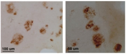
Description Immunohistochemistry (paraffin): at 510ug/ml, positive staining of senile plaques in AD brains.
Handling
Storage This antibody is stable for at least one (1) year at -20°C. Avoid multiple freeze- thaw cycles.
Dilution Instructions Dilute in PBS or medium that is identical to that used in the assay system.
Application Instructions Immunoblotting: at 2-5ug/mL, a band of approx. 70 kDa is detected in HEK293 cell lysate
Immunohistochemistry (paraffin): at 5- 10ug/mL, positive staining of senile plaques in AD brains
These are recommended concen- trations. User should determine optimal concentrations for their applications.
Immunohistochemistry (paraffin): at 5- 10ug/mL, positive staining of senile plaques in AD brains
These are recommended concen- trations. User should determine optimal concentrations for their applications.
References & Data Sheet
Data Sheet  Download PDF Data Sheet
 Download PDF Data Sheet


