


Anti-HIF1α Antibody (34039)
$442.00
| Host | Quantity | Applications | Species Reactivity | Data Sheet | |
|---|---|---|---|---|---|
| Mouse | 100ug | WB,IHC,ICC/IF,ELISA | Human |  |
Overview
Product Name Anti-HIF1α Antibody (34039)
Description Anti-HIF1α Mouse Monoclonal Antibody
Target HIF1α
Species Reactivity Human
Applications WB,IHC,ICC/IF,ELISA
Host Mouse
Clonality Monoclonal
Clone ID ESEE122
Isotype IgG1
Immunogen Recombinant protein corresponding to aa 329-530 of human HIF-1a.
Properties
Form Liquid
Concentration Lot Specific
Formulation PBS, pH 7.4; 50% glycerol, 0.09% sodium azide. Purified by Protein G affinity chromatography.
Buffer Formulation Phosphate Buffered Saline
Buffer pH pH 7.4
Buffer Anti-Microbial 0.09% Sodium Azide
Buffer Cryopreservative 50% Glycerol
Format Purified
Purification Purified by Protein G affinity chromatography
Specificity Information
Specificity This antibody recognizes human, mouse, rat, and bovine HIF-1alpha.
Target Name Hypoxia-inducible factor 1-α
Target ID HIF1α
Uniprot ID Q61221
Alternative Names HIF-1-α, HIF1-α, ARNT-interacting protein
Gene Name Hif1a
Sequence Location Cytoplasm, Nucleus, Nucleus speckle
Biological Function Functions as a master transcriptional regulator of the adaptive response to hypoxia (PubMed:15225651, PubMed:17981124, PubMed:22009797). Under hypoxic conditions, activates the transcription of over 40 genes, including erythropoietin, glucose transporters, glycolytic enzymes, vascular endothelial growth factor, HILPDA, and other genes whose protein products increase oxygen delivery or facilitate metabolic adaptation to hypoxia (PubMed:15225651, PubMed:17981124, PubMed:22009797). Plays an essential role in embryonic vascularization, tumor angiogenesis and pathophysiology of ischemic disease (PubMed:22009797). Heterodimerizes with ARNT; heterodimer binds to core DNA sequence 5'-TACGTG-3' within the hypoxia response element (HRE) of target gene promoters (PubMed:26245371). Activation requires recruitment of transcriptional coactivators such as CREBBP and EP300. Activity is enhanced by interaction with NCOA1 and/or NCOA2. Interaction with redox regulatory protein APEX1 seems to activate CTAD and potentiates activation by NCOA1 and CREBBP. Involved in the axonal distribution and transport of mitochondria in neurons during hypoxia (By similarity). {UniProtKB:Q16665, PubMed:15225651, PubMed:17981124, PubMed:22009797, PubMed:26245371}.
Research Areas Cancer research
Background Hypoxia-inducible factor 1 (HIF-1) is a heterodimeric transcription factor that regulates the transcription of a broad range of genes that facilitate responses to hypoxia including genes that regulate angiogenesis, erythropoiesis, cell cycle, metabolism, and apoptosis. HIF-1 is comprised of two subunits, HIF-1alpha and HIF-1beta. The widely expressed HIF-1alpha is degraded rapidly in normoxic cells by the ubiquitin/proteasomal pathway. HIF-1alpha is proline hydroxylated leading to a conformational change that promotes binding to the von Hippel Lindau protein (VLH)-E3 ligase complex; ubiquitination and proteasomal degradation follows.
Application Images


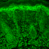
Description Immunohistochemistry analysis using Mouse Anti-HIF1 alpha Monoclonal Antibody, Clone ESEE122 (34039). Tissue: backskin. Species: Mouse. Fixation: Bouin's Fixative and paraffin-embedded. Primary Antibody: Mouse Anti-HIF1 alpha Monoclonal Antibody (34039) at 1:100 for 1 hour at RT. Secondary Antibody: FITC Goat Anti-Mouse (green) at 1:50 for 1 hour at RT. Localization: Membranous and cytoplasmic localization in the epidermis, positive hair follicles, muscle and dermis. .
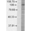
Description Western Blot analysis of Human Cervical cancer cell line (HeLa) lysate showing detection of HIF1 alpha protein using Mouse Anti-HIF1 alpha Monoclonal Antibody, Clone ESEE122 (34039). Load: 15 µg. Block: 1.5% BSA for 30 minutes at RT. Primary Antibody: Mouse Anti-HIF1 alpha Monoclonal Antibody (34039) at 1:500 for 2 hours at RT. Secondary Antibody: Sheep Anti-Mouse IgG: HRP for 1 hour at RT.
Handling
Storage This antibody is stable for at least one (1) year at -20°C.
Dilution Instructions Dilute in PBS or medium that is identical to that used in the assay system.
Application Instructions Immunoblotting: use at 2ug/mL. Predicted molecular weight ~116kDa.This is a recommended concentration.
Endusers should determine optimal concentrations for their applications.
Endusers should determine optimal concentrations for their applications.
References & Data Sheet
References Espinosa I et al. 2008 Am J Surg Pathol 32: 210-218. Lee CH et al. 2010 Adv Anat Pathol 17: 222-232. Not for use in diagnostic or therapeutic procedures.
Data Sheet  Download PDF Data Sheet
Download PDF Data Sheet